Breast examination without radiation; if needed, the doctor also checks the armpit lymph nodes. Does not replace a mammogram if one is recommended for you.
Worth having if:
•a lump or change in breast shape
•tenderness, nipple discharge
•dense breast tissue — as a complement to mammography
•referral from a breast specialist or gynecologist
Certified Grand Medical specialists. Each doctor has their own types of examinations. If you are not sure whom to book with, we will help you choose a specialist.
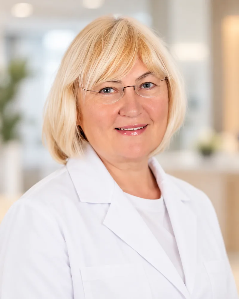
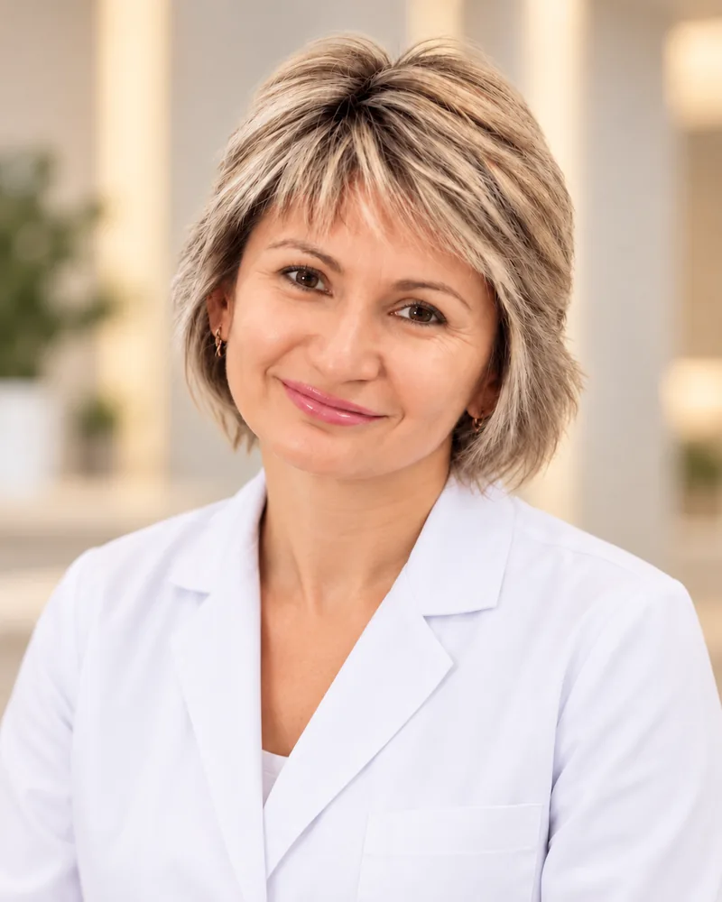
Alla Khachaturyants
Ultrasound doctor
Ultrasound doctor with many years of experience. Specializes in breast ultrasound.
Ultrasound, Doppler and echocardiography — what's the difference
All three methods use ultrasound and involve no radiation, but they answer different questions: what an organ looks like, how blood moves through vessels and how the heart works.
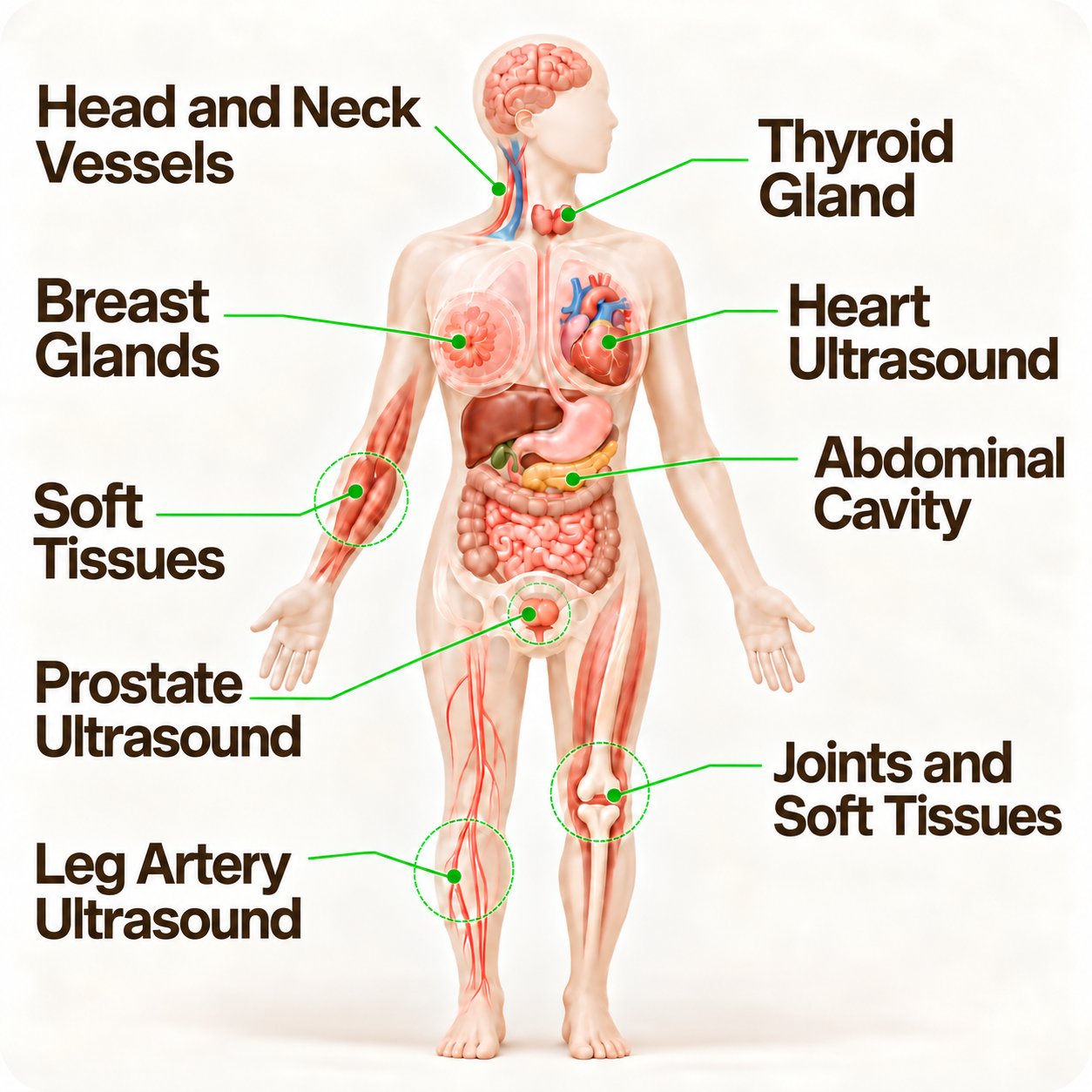
Organ and tissue ultrasound
Classic ultrasound examination: a real-time image of the organ. The doctor assesses the size, shape and structure of tissues and describes any changes found.
Used for:
•thyroid and breasts
•abdominal organs and kidneys
•prostate
•joints, muscles and soft tissues
Good to know: Ultrasound helps detect changes and decide on next steps, but it does not replace a consultation with a specialist or other methods — mammography, CT, MRI or biopsy, where needed.
Doppler and duplex scanning
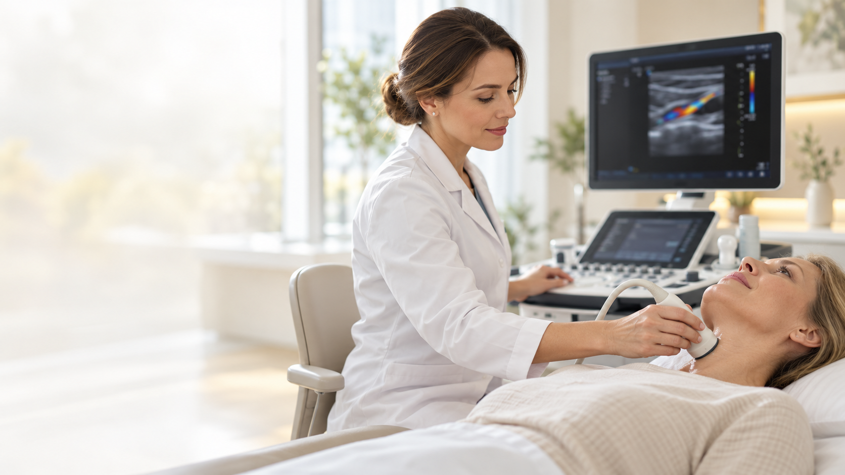
Doppler shows the movement of blood. Duplex scanning combines an image of the vessel itself with an assessment of blood flow in it — in the arteries of the neck, arms and legs, and in veins.
What the doctor assesses:
•the state of the artery wall and plaques
•degree of narrowing (stenosis)
•speed and direction of blood flow
•vein patency, valve function, clots
Good to know: narrowing of the carotid arteries is one of the risk factors for stroke. If it is found, the treating doctor can arrange prevention or treatment in good time.
Echocardiography
Echocardiography shows the heart in motion: how its walls contract, how the valves open and close and how blood passes through the heart chambers.
What the doctor assesses:
•the size of the heart chambers
•ventricular wall thickness
•contractility and ejection fraction
•valve function and blood flow inside the heart
Good to know: echocardiography is usually ordered by a cardiologist or family doctor — for example, for shortness of breath, a heart murmur, high blood pressure or before surgery.
When to have an ultrasound
Many changes stay silent for a long time
On a doctor's referral. Ultrasound is most often ordered by a family doctor, endocrinologist, gynecologist, urologist, cardiologist or neurologist — to clarify the cause of symptoms or check changes in blood tests.
No symptoms — based on individual indications. Whether you need preventive tests and how often depends on age, family history and risk factors. It is best to discuss this with your doctor.
No radiation. Ultrasound uses no X-rays, so it can be repeated for monitoring when needed.
Follow-up over time. If a nodule, cyst or plaque has already been found, a repeat ultrasound helps the doctor see whether it changes over time.
0
radiation
No X-ray exposure
1
visit
Examination, report and images right away
9
areas
Ultrasound of organs, heart, vessels and tissues
Good to know
You can book without a doctor's referral
We work with the insurers BTA, Balta and Seesam — please check the terms in your policy
Several examinations can be done in one visit — by prior arrangement
If needed, we will refer you to a clinic specialist
Painless and quick
How an ultrasound goes
1
Booking
Choose an examination and a convenient time online or by phone.
2
Preparation
Some examinations require preparation — for example, abdominal ultrasound is done on an empty stomach. You will learn everything you need when you book.
3
Examination
The doctor moves a probe over the skin with a little gel and assesses the image. It is painless.
4
Report
Right after the examination you receive the doctor’s report and printed images.
Modern equipment
Modern equipment for accurate diagnostics
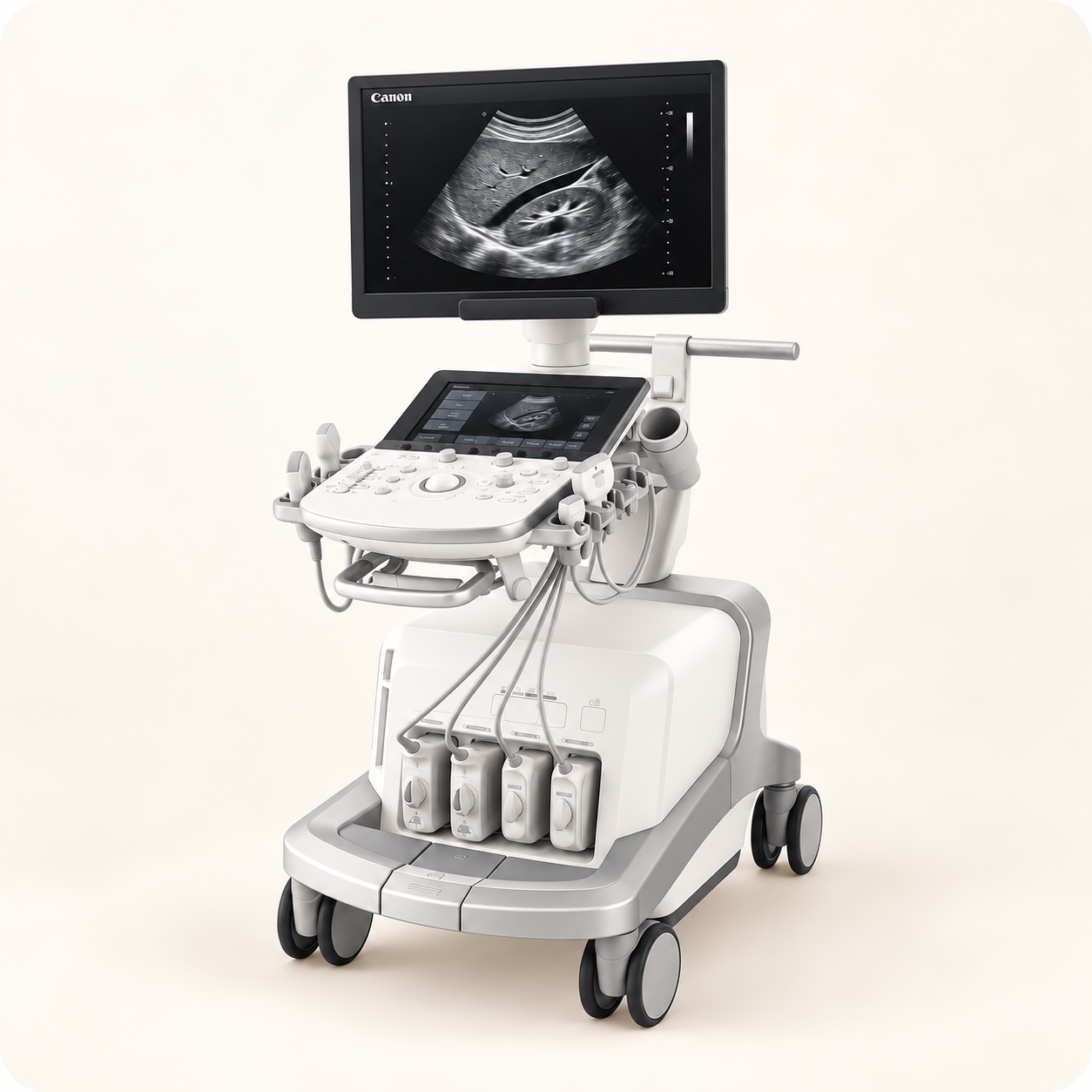
At Grand Medical, ultrasound examinations are performed on Canon Aplio i900 and GE Vivid T8 systems. Modern imaging and Doppler technology helps the doctor assess organs, tissues and blood flow in detail.
Canon Aplio i900 — an expert-class system for internal organs, vessels and superficial tissues
GE Vivid T8 — a system for echocardiography: assessing the structure and function of the heart
Linear, convex and sector probes — for superficial organs, the abdomen and the heart
I was pleasantly surprised how quickly I could book — especially since I was in Latvia for only a couple of days. Very convenient.
Olga KravaleGoogle review · translated
I have had ultrasounds in many places, but I have never met such an attentive and professional approach. Everything was explained, questions were answered, and you can feel the care for the patient.
Angel ShopGoogle review · translated
Booking
Book an ultrasound
The quickest way is the booking portal: you can see the doctors’ free slots and pay straight away. Not sure which examination to choose? Leave a request or call us — we’ll help you choose and book.
Ļauj atklāt aterosklerozi, trombus, aneirismas un citas patoloģijas. Tiek izmantota slimību diagnosticēšanai, kā arī profilaktiskajiem izmeklējumiem, īpaši cilvēkiem ar riska faktoriem.
 Expert-class equipment
 Experienced doctors