
Heart ultrasound · Riga, Purvciems

Echocardiography in Riga — heart ultrasound

Echocardiography (echo) helps assess the structure and function of the heart: chamber size, valve condition, contractility and blood flow. The examination uses ultrasound, with no X-ray radiation.

  • No referral needed
  • Report on the day of the examination
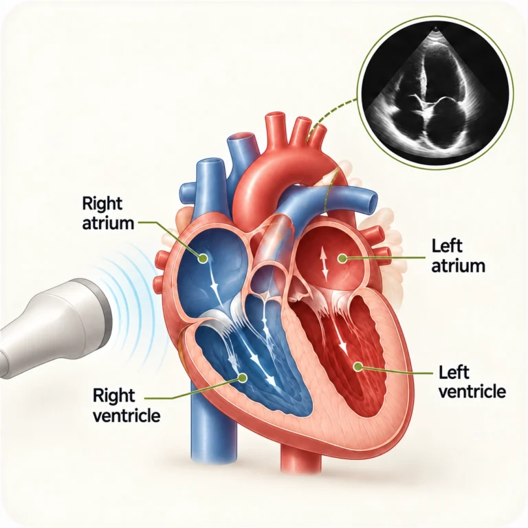
Diagram of the heart chambers with an echo image: right and left atria, right and left ventricles

Indications

When a doctor may recommend an echo

Whether you need the examination is decided by a doctor. An echo is most often recommended in these situations:

  • Shortness of breath, swelling and reduced exercise toleranceAn echo helps assess whether symptoms may be linked to changes in the structure or function of the heart.
  • Heart murmurs and ECG changesThe examination allows the doctor to assess the heart valves, chambers and contractility.
  • Monitoring of heart conditionsFollow-up in heart failure, valve disease and other diagnosed conditions.
  • Follow-up after illness or proceduresYour doctor may recommend an echo to see how the heart is working over time.

Sudden severe chest pain, sudden shortness of breath or loss of consciousness — do not wait for a routine appointment, call 113 or 112 immediately.

What the doctor assesses

What echocardiography shows

Ultrasound shows the heart in motion. During the examination the doctor assesses:

  • Icon: heart chamber size

    Size of the heart chambers

    The doctor measures the atria and ventricles and looks for any changes in their size.

  • Icon: heart wall thickness and motion

    Wall thickness and motion

    The examination shows the thickness of the heart muscle and how it contracts.

  • Icon: heart pumping function

    Pumping function

    It shows how effectively the heart pumps blood, including the ejection fraction.

  • Icon: heart valve function

    Valve function

    The doctor checks how the valves move and looks for narrowing or backward flow.

  • Icon: blood flow in the heart

    Blood flow in the heart

    Doppler imaging helps assess the direction and speed of blood flow.

  • Icon: fluid around the heart

    Fluid around the heart

    An echo can detect fluid collecting in the pericardium.
Who performs the examination

Doctors who perform echocardiography

The examination is performed by the clinic’s cardiologists. Our reception will help you choose a doctor and a convenient time.

Елена Шафро / Elena Shafro / Jeļena Šafro

Elena Shafro

Certified cardiologist

Cardiology consultations and echocardiography for adults, including Doppler measurements.

Doctor’s profile →

Галина Блументале / Galina Blumentale / Gaļina Blūmentāle

Galina Blumentale

Cardiologist

Echocardiography (echo) for adults.

Doctor’s profile →

Виталий Гребёнкин / Vitaly Grebyonkin / Vitālijs Grebjonkins

Vitaly Grebyonkin

Certified cardiologist

Echocardiography specialist, interventional cardiologist.

Doctor’s profile →

Step by step

How an echo works and how to prepare

A standard echo: the probe is placed on the skin of the chest. The appointment takes about 15 minutes.

  1. Booking

    Online, by phone or on WhatsApp. No referral needed.

  2. Talk with the doctor

    The doctor asks about the reason for the examination and reviews previous results.

  3. Ultrasound examination

    You lie on a couch; the doctor applies gel and moves the probe over your chest.

  4. Report

    You receive the diagnostic report on the day of the examination.

Preparing for the examination

  • No special preparation is usually needed, and you do not need to fast.
  • Take your prescribed medicines as usual unless your doctor has advised otherwise.
  • Bring previous echo and other heart test results, if you have them.
  • Wear clothes that are easy to remove to the waist.

What you will feel

The examination is painless: you may only notice the cool gel and light pressure from the probe. You can return to your usual activities straight away.

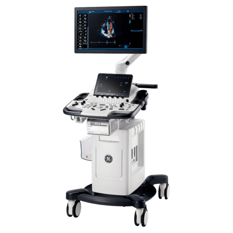
GE Vivid T8 ultrasound system — photo of the model

Equipment

Examination on a GE Vivid T8 system

At Grand Medical, echocardiography is performed on a GE Vivid T8 ultrasound system that shows the heart in real time. The doctor assesses wall motion, valve function and blood flow using Doppler modes.

Price

Price and insurance

Echocardiography (echo)

85 €

Heart ultrasound examination with a diagnostic report. No referral is needed to book.

An echo and a cardiology consultation are separate services. If you need your results explained, treatment advice or further tests, you can book a cardiologist consultation (paid separately).

Health insurance

  • Balta
  • Seesam
  • BTA

The clinic works with Balta, Seesam and BTA. Cover depends on the terms of your policy — please check with your insurer before your visit.

Diagnostic methods

Echo, ECG and Holter — a quick comparison

These tests complement each other — your doctor decides which one you need.

  • Echo (heart ultrasound)

    Shows the structure of the heart, the valves, wall contraction and blood flow.

  • ECG

    Records the heart's electrical activity and helps assess rhythm and conduction.

  • Holter

    Records the ECG over a longer period to detect rhythm problems that a standard ECG may miss.

FAQ

Frequently asked questions

Yes. Echocardiography is an ultrasound examination of the heart, also called a heart ultrasound or echo.

An echo shows images of the heart and how it works, while an ECG records electrical activity and rhythm. See the comparison.

No, you can book without a referral.

Usually not: you do not need to fast and you take your medicines as usual. Bring previous results if you have them.

On the day of the examination.

No. The €85 price covers the examination and the diagnostic report. A cardiologist consultation is a separate service.

The clinic works with Balta, Seesam and BTA. Cover depends on your policy — please check with your insurer.

Booking

Book an echo

Booking portal, phone, WhatsApp or a request on our website — choose what suits you.

Book online

If there is no suitable time on the booking portals, contact the clinic — we will help you find an available slot.

By sending a request, you agree to our personal data processing policy.

Galvas un kakla asinsvadu doplerogrāfija (DUPLEX)

1 novembrī

Ļauj atklāt aterosklerozi, trombus, aneirismas un citas patoloģijas. Tiek izmantota slimību diagnosticēšanai, kā arī profilaktiskajiem izmeklējumiem, īpaši cilvēkiem ar riska faktoriem.