
For pain, swelling and after injuries. The doctor assesses joints, muscles, tendons, ligaments and surrounding soft tissues, including during movement. This examination is called MSK ultrasound — musculoskeletal ultrasound.
Joint or soft tissue ultrasound price €70
Patients with Balta, Seesam and BTA policies are welcome.
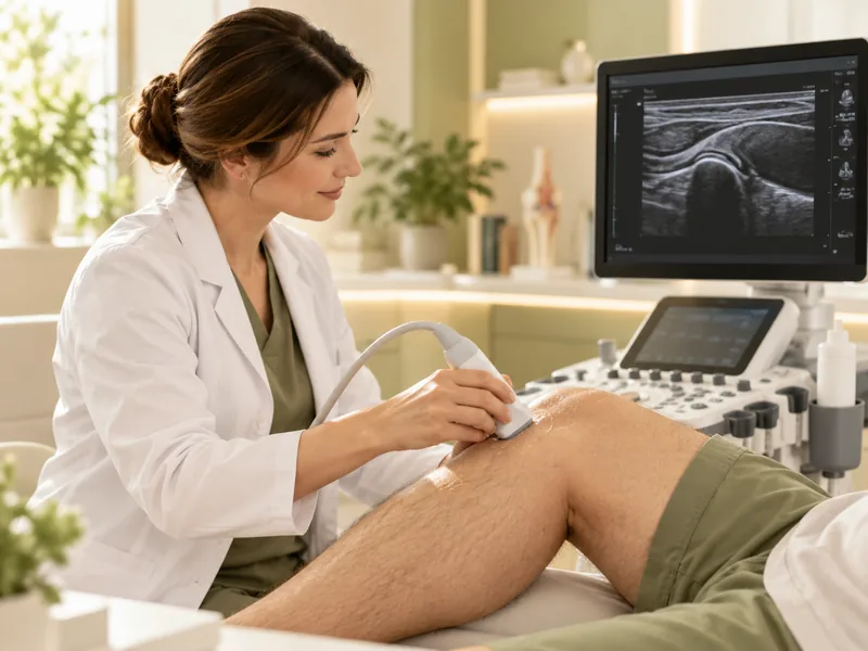
Assessing tendons and ligaments while bending and turning
Depending on the area and number of zones
Ultrasound without ionising radiation
Stirnu iela 8, Valeo medical centre
The examination helps clarify the cause of complaints in the joints, muscles and surrounding soft tissues, and assess changes after injuries.
Joint pain, limited mobility, a feeling of tightness; assessment of soft tissue changes in osteoarthritis.
Sprains, partial tears, suspected tendon and muscle ruptures, sports injuries.
Synovitis, bursitis, joint effusion; rheumatoid arthritis, gout, spondyloarthritis.
Tendinitis, tenosynovitis, enthesopathies, tenderness at attachment points.
Signs of peripheral nerve compression or inflammation — entrapment syndromes.
Cysts, lipomas, fibromas, enlarged lymph nodes, hernias and other soft tissue masses.
MSK ultrasound is also used to monitor treatment of chronic joint conditions, before surgery and for follow-up afterwards — as referred by a doctor.
We examine joints and surrounding tissues that may be linked to pain, swelling or limited movement. A key advantage of MSK ultrasound is that the doctor sees structures in motion, which X-ray cannot do.
The area is chosen based on your complaints and the purpose of the examination.
The doctor examines tissue structure and signs of damage and inflammation.
The examination usually takes 15–30 minutes. The doctor may ask you to bend or turn the limb to assess structures in motion.
Special preparation is usually not requiredWear comfortable clothing with access to the area. If you have previous MRI, X-ray or ultrasound results, bring them with you.
The doctor clarifies your complaints and gets access to the area. Sitting or lying — depends on the joint.
Gel is applied to the skin and the tissues are examined with the probe, in motion if needed (functional tests).
The doctor explains the results right after the examination; the report is usually ready on the day of the visit.
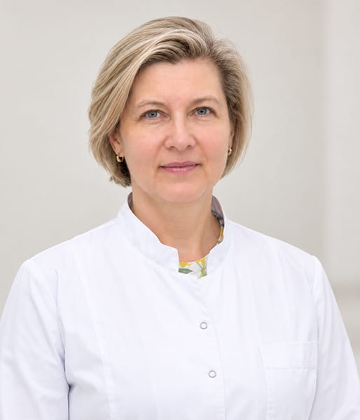
Internist, ultrasound specialist
A certified doctor with many years of experience in ultrasound examinations. Specialises in ultrasound of muscles, tendons, joints and bone structures.
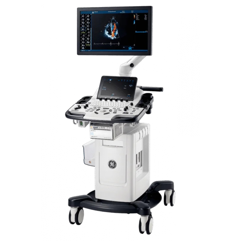
Equipment
The examination is performed on an expert-class General Electric Vivid T8 system. For precise detail of small joints and tendons, a special GE L8-18i mini-probe (“hockey stick”) is used.
Grand Medical also operates a Canon Aplio i900 ultrasound system.
I was pleasantly surprised how quickly I was able to book — especially considering I had only been in Latvia for a couple of days. Very convenient.
I have had ultrasound scans in many places, but I have never met such an attentive and professional approach. They explained everything, answered my questions, and you can feel that they care about the patient.
Answers to questions that often come up before the examination.
No, the examination is painless and non-invasive. You may feel light pressure from the probe over a tender area.
Not always — the methods complement each other. Ultrasound works well for superficial structures and assessing them in motion, MRI for deeper tissues and cartilage. The choice depends on your complaints and the doctor’s recommendation.
Usually not. Comfortable clothing that lets you uncover the area is enough.
The doctor gives a preliminary report immediately and the final one on the day of the examination. In complex cases the doctor will agree on a timeframe.
The fastest way is through a booking portal, where you can see doctors’ available times. If you are unsure which specialist to see, leave a request or call us — we will help you choose and book you in.
Online booking
Free appointment times with doctors are available on the booking portals. If you didn’t find a suitable time, contact us — we will help find a solution.
If the ultrasound reveals changes, the doctor will advise which specialist to see.

Ļauj atklāt aterosklerozi, trombus, aneirismas un citas patoloģijas. Tiek izmantota slimību diagnosticēšanai, kā arī profilaktiskajiem izmeklējumiem, īpaši cilvēkiem ar riska faktoriem.