
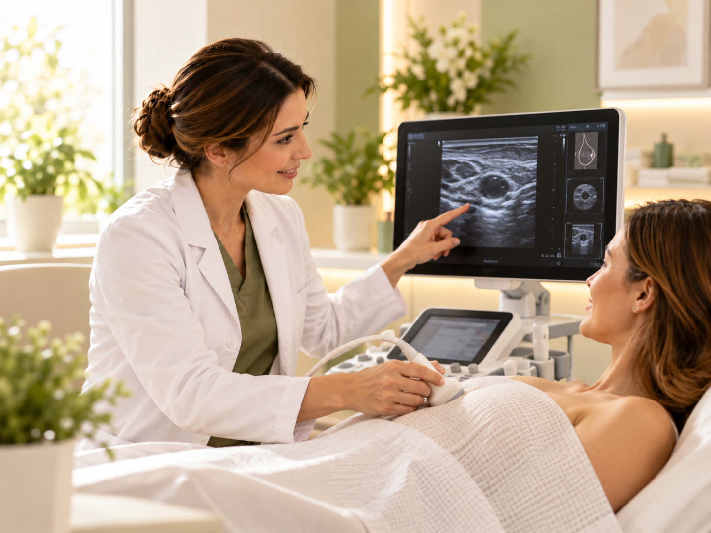
Breast ultrasound helps assess the condition of breast tissue and examine lumps, cysts and other changes. The examination is performed by experienced Grand Medical specialists in Riga, Purvciems. If needed, the doctor also assesses the armpit areas and explains the results.
Breast ultrasound €75
With Dr. Alla Khachaturyants — €90. Patients with Balta, Seesam and BTA policies are welcome; coverage depends on your policy terms.
And the armpit areas — if needed
Depending on the scope of the examination
Ultrasound is suitable during pregnancy too
Stirnu iela 8, Valeo medical centre
Breast ultrasound is performed when you have symptoms, to clarify the results of other examinations and to monitor known changes. No referral is needed.
A new lump or area of tissue that feels different from the surrounding structures.
Local tenderness, unpleasant sensations or changes in a particular area of the breast.
Unusual discharge, especially if it appears spontaneously or contains blood, needs timely medical assessment.
A change in breast shape, nipple retraction, local swelling or visible skin changes.
Follow-up of previously found cysts, fibroadenomas and other changes as recommended by a doctor.
Assessment of post-operative changes, tissue around implants or symptoms that need further diagnostics.
Breast ultrasound may also be recommended to clarify the results of other examinations and to monitor previously detected changes. The choice of examination and the need for repeat scans are decided individually.
Ultrasound is well suited to examining the soft tissues of the breast. It helps distinguish a fluid-filled lesion from a solid one and assess the shape, margins and size of changes, as well as their position relative to nearby tissue.
The doctor decides the scope of the examination based on your symptoms and its purpose.
The doctor describes any findings in the report.
Ultrasound allows the doctor to assess tissue structure in real time and determine the location and size of any changes. When monitoring over time, the results can be compared with previous examinations. On its own, ultrasound neither confirms nor rules out a malignant disease: if the findings are not sufficient, the doctor may recommend further diagnostics.
Breast ultrasound and mammography are different imaging methods that can complement each other. Ultrasound uses sound waves and helps assess tissue structure: examining lumps that have been found, telling a cyst from a solid lesion and clarifying the nature of some changes. It involves no radiation, so it can be performed during pregnancy and breastfeeding.
Mammography uses X-rays and plays an important role in breast cancer screening. The choice of method depends on age, symptoms, individual risk and previous results.
Ultrasound does not replace a prescribed mammogramIf your doctor has recommended mammography, it should not be replaced by ultrasound alone.
The examination usually takes 15–20 minutes. The doctor works step by step and answers your questions during the visit.
Preparing for a breast ultrasoundSpecial preparation is usually not needed. If you have previous ultrasound, mammography or MRI results or medical reports, bring them with you — this helps the doctor assess changes over time.
The doctor clarifies your symptoms and reviews any previous results. You lie on the couch and uncover the breast area for the examination.
A special gel is applied to the skin. The doctor systematically examines the tissue of both breasts with the ultrasound probe and, if needed, the armpit areas.
After the examination the doctor explains the results. If changes are found that need further assessment, the specialist recommends the next steps.
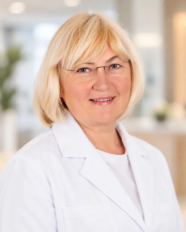
Breast ultrasound doctor
A doctor with many years of experience, previously at RAKUS Gaiļezers. Breast ultrasound is her main field. Sees patients in Latvian and Russian.
Breast ultrasound — €90
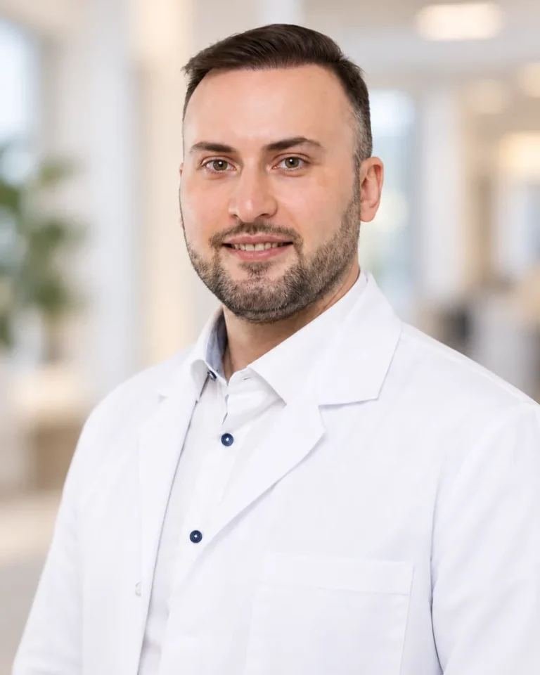
Ultrasound doctor
A doctor with extensive experience in diagnostic radiology who headed a radiology department at Riga 1st Hospital. Performs breast, thyroid and abdominal ultrasound.
Breast ultrasound — €75

Obstetrician-gynecologist, ultrasound doctor
Obstetrician-gynecologist and ultrasound doctor with 25+ years of experience. Gynecology consultations, breast, thyroid, abdominal and prostate ultrasound.
Breast ultrasound — €75
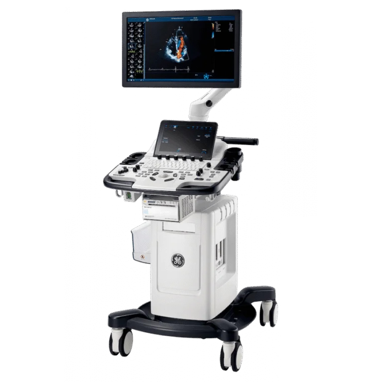
Equipment
Breast ultrasound at Grand Medical is performed on expert-class ultrasound systems Canon Aplio i900 and GE Vivid T8.
I am very pleased with my visit to the Grand Medical clinic. Experienced specialists work here, all in one place, and I especially want to highlight Alla Khachaturyants. Thank you! I recommend this clinic to everyone.
I have had ultrasound scans in many places, but I have never met such an attentive and professional approach. They explained everything, answered my questions, and you can feel that they care about the patient.
Answers to the questions patients most often ask before the examination.
If you still have menstrual cycles, the doctor may recommend the first half of the cycle for a planned examination: at this time breast tissue is usually less tense and sensitive, and the image is easier to assess. There is no strict rule for everyone. If you are worried about a lump, pain or discharge, do not postpone the examination to wait for a particular day — book the nearest convenient time.
No special preparation is required: you do not need to follow a diet or come on an empty stomach. It is more convenient to wear clothing that is easy to remove to the waist. Bring the results of previous ultrasound, mammography or MRI examinations and medical reports — the doctor will be able to compare the data and assess changes over time.
The examination is usually well tolerated and painless. The doctor applies gel to the skin and moves the probe smoothly. If the breast is tender or inflamed, the pressure of the probe may cause slight discomfort — tell the doctor, and they will work as gently as possible.
Yes. Ultrasound does not use ionising radiation, so it can be performed during pregnancy and breastfeeding when there is a medical reason — for example, a new lump or pain. If you are breastfeeding, it is more convenient to come for the examination shortly after feeding or pumping.
There is no universal recommendation for all women to have a breast ultrasound every year. The doctor decides how often you need examinations based on your age, symptoms, family history and previous results. For example, when monitoring a cyst or fibroadenoma, the doctor may schedule a follow-up ultrasound after a certain period.
No. They are different methods that often complement each other. Ultrasound shows the structure of dense tissue well and helps clarify the nature of a lump, while mammography remains the main method of breast cancer screening. If mammography is indicated for you, ultrasound alone is not a full replacement.
The fastest way is through a booking portal, where you can see doctors’ available times. If you are unsure which examination to choose, leave a request or call us — we will help you decide and book you in.
Online booking
Can’t find a suitable time, or are all slots taken? Contact us — we’ll help you book an appointment.
Grand Medical also performs thyroid, abdominal, joint and soft tissue ultrasound.

Ļauj atklāt aterosklerozi, trombus, aneirismas un citas patoloģijas. Tiek izmantota slimību diagnosticēšanai, kā arī profilaktiskajiem izmeklējumiem, īpaši cilvēkiem ar riska faktoriem.